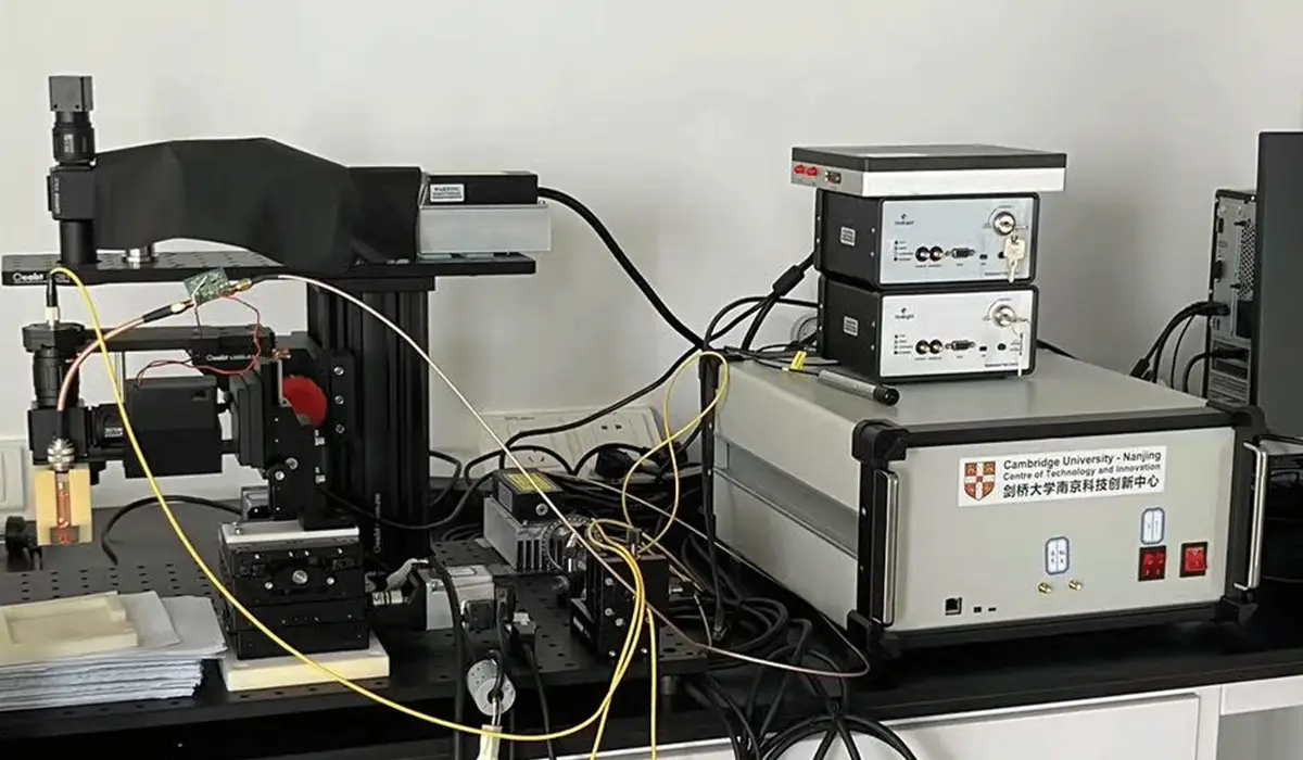
Dual-band photoacoustic microscope prototype
Prototype/results show · 2026/11/5 · 11 次浏览
The dual-band photoacoustic microscope prototype developed in this project, using specific wavelength laser to irradiate the tissue, the optical contrast between different tissues in the photoacoustic
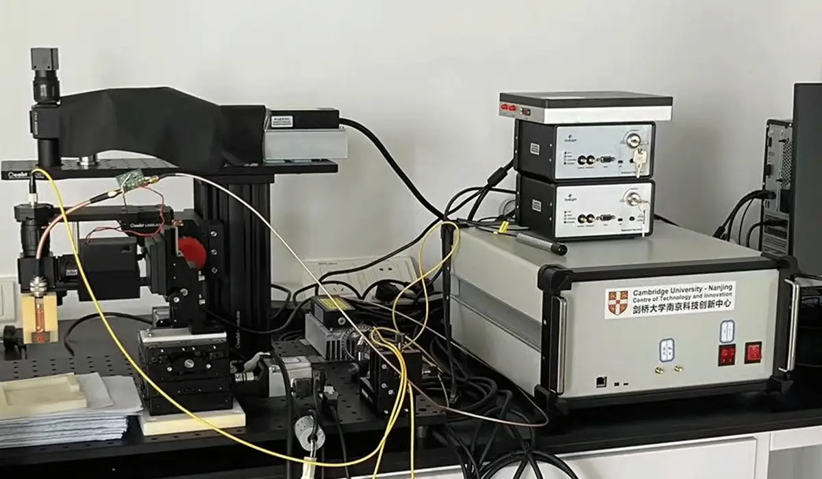
The dual-band photoacoustic microscope prototype developed in this project, using specific wavelength laser to irradiate the tissue, the optical contrast between different tissues in the photoacoustic image is high, which can realize the structure and function imaging of biological tissue in the unlabeled state, which provides an important means for the study of the morphological structure, physiological characteristics, pathological characteristics, metabolic function of biological tissue. The main advantage of photoacoustic imaging over traditional medical imaging techniques is that it is a non-invasive, non-ionizing, non-invasive imaging technique that can be used for in-vivo detection with 100 percent sensitivity and therefore can also provide quantitative information.
dual-band photoacoustic microscope prototype is equipped with laser heads with 266nm and 532nm wavelengths. The specific technical information is as follows:
· Imaging Resolution:532nm is about 1.8 μm, and the resolution of 266nm is about 1.3 μm.
·imaging depth:In vivo experiments demonstrated that microvessels under the skin can be imaged within a depth of 1mm.
·imaging range:is about 40 × 40mm2, and the imaging results of the phantoms show that the prototype achieves accurate imaging in the range of 15 × 15mm2, and the image reconstruction results are consistent with the actual object structure.
application prospect
♦ DNA/RNA has strong light absorption in the ultraviolet region (180-400 nm). Photoacoustic microscopy enables cell nucleus imaging by using a laser source with a wavelength of 266 nm. Since the canceration of cells usually leads to great changes in the morphology and density of cells, the imaging of the nucleus is expected to provide a reference for the early diagnosis of cancer in clinic. In addition, compared with the traditional hematoxylin and eosin staining methods, this method does not require staining and is conducive to real-time pathological evaluation of intraoperative sliced tissues.
The prototype is based on the reflection mode and is suitable for untreated fresh thick tissue, which can further shorten the evaluation time. The blood circulation system is one of the most important physiological systems. Hemoglobin is the most abundant protein in the blood and an important oxygen carrier, which is closely related to metabolism. Imaging of hemoglobin can reflect whether physiological conditions associated with the circulation are abnormal. Hemoglobin has strong light absorption in the visible region (400-700nm). The use of 532nm wavelength photoacoustic microscope can be used for high-contrast imaging of microvascular network, which is expected to play a role in the field of cerebral blood flow monitoring, diagnosis and treatment of ophthalmic diseases, skin vascular related pathology research and so on.
In the exploration of physiological section detection in this project, based on the performance characteristics of photoacoustic microscope such as no staining (fast speed, avoiding potential artifacts and sample damage caused by dyes) and realizing high-resolution and high-contrast imaging, it has been proved that photoacoustic microscope can simultaneously display the nucleus and the density of cells, reflecting the heterogeneous nature of tumor nuclei to a certain extent.